Diabetic retinopathy’s advanced stages trigger abnormal and fragile new blood vessels to form, leading to vision loss or even blindness.
As early signs of retinopathy are often not noticeable, it is vitally important that regular eye exams with dilation occur.
Blurred Vision
Blurred vision refers to any loss of sharpness of visual imagery. This condition may arise in either eye, and be indicative of numerous eye diseases and disorders. Aging also causes blurred vision to worsen over time and requires glasses or contact lenses as treatment – though, in rare cases, blurred vision may signal something more serious is happening in both eyes.
Blurry vision may be caused by conditions that affect the ocular surface (the front “window” of the eye). These include dry eyes, which is often an early warning sign of corneal scarring or cataracts; as well as signs of glaucoma and some neurological diseases. It’s important to differentiate between occasional blurriness that comes and goes from age-related blurriness as well as conditions like astigmatism or hypermetropia that gradually worsen over time.
Diabetes-induced retinal damage causes blurry vision due to damage to retinal blood vessels that over time become damaged and leak fluid into macular edema (swelling). Over time, this leakage of fluid into the retina leads to macular edema (swelling). Over time this fluid may even result in permanent loss of central vision.
Other symptoms of diabetic retinopathy can include losing the ability to see colors, dark areas in your vision known as scotoma and seeing floaters. Over time these symptoms can develop, making it harder than ever to notice without regular eye exams; with careful control and regular visits to an ophthalmologist can prevent or delay these from emerging.
Your doctor can conduct various eye health exams to evaluate your eye health, such as an examination with dilated pupils. At this exam, your doctor can look for fluid leakage from diabetic retinopathy’s damaged blood vessels as well as take photos using fluorescein angiography or optical coherence tomography angiography of the retina to help assess its severity. Your physician can then suggest the most suitable treatments options and suggest tissue plasminogen activator (tPA). In rare cases, they may use TPA to treat conditions that cause blurred vision.
Spots or Floaters
Floaters are small clumps of gel or cells found within your vitreous, which is the jelly-like substance located at the back of your eye, that cast shadows onto the retina and appear as dots, rings, lines or cobwebs in your vision. Although distracting at times, floaters typically resolve over time without any permanent damage being done to your vision; however if they suddenly appear in large numbers or accompany flashes of light they should be evaluated by a retina specialist as this could indicate posterior vitreous detachment (PVD).
PVD, or vitreous vitreous detachment disease, is a common side effect of aging that occurs when the vitreous shrinks gradually away from its original position in the retina’s inner lining, eventually becoming detached. While symptoms usually do not arise initially, PVD could eventually lead to retinal tear formation which eventually develops into detachment requiring treatment by retina specialists and thus anyone experiencing new floaters and flashes of light should have their eyes examined to rule out more serious problems such as PVD before treating any symptoms may ensue.
Sometimes vitreous bleeding can also result in floaters. This occurs more frequently when there are complications related to diabetes or conditions that affect blood vessels; an extremely rare genetic condition known as Ehlers-Danlos Syndrome may also contribute to this form of bleeding.
When experiencing these symptoms, medical assistance should always be sought immediately. If floaters or flashes are combined with any other symptoms, consider it an emergency and get to the nearest hospital immediately; early treatment increases your chance of recovering your vision due to retinal detachment which requires specialist treatment from eye hospitals; left untreated, retinal detachments may lead to severe blindness – but with early treatment they’re usually preventable; their signs often resemble those of PVD and should be treated as urgent eye emergencies.
Difficulty Seeing at Night
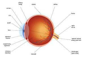
The retina is a layer of cells lining the backwall inside your eye that senses light and sends signals to your brain so you can see. It is especially crucial that the center area (called macula) works properly; otherwise you may experience trouble seeing fine details or different colors; difficulty seeing at night may also be a symptom of diabetic retinopathy, one of the leading causes of blindness among American adults aged 25 to 65 years.
Diabetic retinopathy occurs when elevated blood sugar levels damage the tiny blood vessels supplying your retina, damaging its supply. Early signs may not appear; over time however, weakened vessels begin leaking fluid into the retina, clouding central vision (macular edema).
As the disease advances, new blood vessels form on the retina’s surface. Unfortunately, these abnormal vessels can easily rupture and bleed easily, blocking oxygen flow to the retina and leading to further loss of vision. Furthermore, as blood vessels continue to bleed they may eventually dislodge from it altogether either gradually over time or suddenly.
Diabetes patients should undergo a dilated eye exam at least annually to detect early signs of retinopathy and reduce the risk of vision loss or blindness. They should also follow a healthy diet, exercise regularly and take their medications as directed in order to keep their blood sugar under control.
If you experience blurred or distorted vision, spots or floaters, or difficulty seeing at night, consult your eye doctor immediately. Your physician can give instructions to keep your blood sugar under control to avoid retinal detachment and other eye complications.
An eye exam involves dilatation of your pupils for examination of your retina and its supporting structures, along with questions regarding past health history and family relationships. Your physician may use other tests like optical coherence tomography (OCT) or fluorescein angiography to check your retina for damage or abnormal new blood vessels.
Loss of Vision
Diabetics at risk for diabetic retinopathy can become increasingly likely over time as their disease worsens, with blood vessels that supply oxygenated blood to the retina becoming damaged and possibly swelling or leaking fluid, eventually resulting in blurred vision or even blindness as the disease advances.
Nearly half of American’s with diabetes have some form of retinopathy. Non-proliferative diabetic retinopathy (NPDR), the most prevalent form, starts as tiny blood vessel leaks that progressively get worse until they lead to fluid accumulation in the macula and macular edema occurs. Diabetes mellitus is the leading cause of blindness among American adults aged 25-65 and its most serious form is proliferative diabetic retinopathy or PDR – caused by abnormal new blood vessels growing on the retinal surface. These vessels can bleed into the eye, leading to vitreous hemorrhage. Furthermore, the leakage from these blood vessels may also cause new scar tissue formation on retina, pulling away from its supporting tissues and leading to detached retinas.
Your risk of vision loss can be decreased by maintaining stable blood sugar levels, receiving regular dilated eye exams and following medical advice from your doctor. If symptoms arise, visit an ophthalmologist immediately as early treatment may increase chances of recovering lost vision.
Your ophthalmologist may use several tests to screen for diabetic retinopathy, including fundus photography and optical coherence tomography (OCT). Drops will be placed into your eye to dilate it so the doctor can look inside with a special lens. Fluorescein angiography or OCT angiography may also be performed to show retinal blood vessel condition.
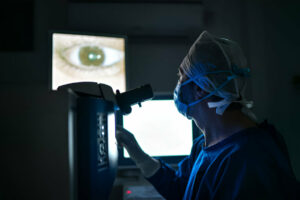
Laser surgery allows your ophthalmologist to make several hundred tiny burns on the retina to seal off leaky blood vessels and stop their growth, as well as shrink abnormal ones and reduce macular swelling. If retinopathy is advanced, they might recommend vitrectomy surgery, in which your surgeon removes vitreous gel and blood from leaking vessels in the back of the eye.


