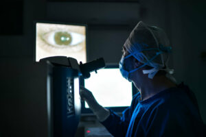
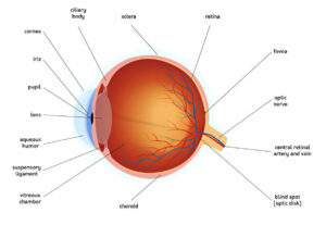
At cataract surgery, drops are used to dilate your pupil. After creating an incision and using ultrasound energy to break apart your cataracts, they are then suctioned out through an outpatient surgery process.
Your surgeon will then replace the cataract with an intraocular lens implant (IOL), enabling light to pass through and focus properly onto your retina.
Infection
Infection is a potential threat following cataract surgery, and should be considered carefully when making decisions pertaining to surgery. It could impact either ocular structures or surrounding tissues and could be caused by bacteria, fungus or virus infections. While such instances are relatively rare, it’s still important to understand their risk and take necessary precautions against infection.
Before cataract surgery, your ophthalmologist will perform a preliminary eye exam to make sure you’re suitable. If this is determined to be true, they’ll schedule the operation and create an incision in your cornea before inserting a small instrument to break up cloudy lens fragments; after which it’s suctioned off and replaced by an intraocular lens implant (IOL) that corrects vision problems caused by cataracts.
Following surgery, your doctor may provide several eye drops to protect you from infection and regulate eye pressure. These may include antibiotics like gatifloxacin or gentamicin as well as steroids to reduce inflammation or lubricating drops to ease dryness in the eye. Diabetics or those suffering from conditions that compromise immunity may be at a greater risk for infections;
Endophthalmitis is one of the main risks associated with cataract surgery. It’s a sudden and severe infection of the vitreous humor (the center-point fluid that fills up most of your eye). This could be caused by infection, injury, penetrating trauma or drug reactions; typically staphylococcal or streptococcal bacteria are to blame but fungal or viral/parasitic diseases like herpes simplex virus and lupus may also play a part in its formation.
Patients diagnosed with infectious endophthalmitis typically do better than those who receive noninfectious endophthalmitis; however, outcomes in cases involving trauma involving gram-negative organisms tend to be less favorable. Precautions you can take after cataract surgery to minimize infection risks include washing your hands before applying eye drops and not touching the eye drop bottle directly.
Disruption of the Posterior Capsular Bag
Under cataract surgery, your doctor will perform an outpatient procedure in which they remove and replace your natural lens with an artificial one in order to restore vision. Typically this takes around an hour; during which eye drops may be used to numb your eye as well as possibly be given as a sedative medication.
Cataracts typically develop gradually and may remain undetected until they begin interfering with your vision. You will typically become aware of cataracts when they interfere with daily tasks like reading or driving; cataracts can make your vision appear cloudy, making it hard to clearly see nearby images as well as those further away.
Phacoemulsification is an increasingly popular procedure to remove cataracts. Your surgeon makes a tiny incision in your cornea and inserts an instrument which breaks up the center of the cataract into microscopic particles that can then be suctioned away by suctioning machines. Finally, they insert an intraocular implant crafted from plastic, silicone or acrylic so light can pass freely through and focus directly onto your retina for clear vision restoration.
However, cataracts may recur and your vision deteriorate after cataract surgery requiring further procedures to restore vision. One common side effect associated with surgery is posterior capsule opacification (PCO), whereby your lens capsule becomes cloudy impairing vision and leading to further procedures being required to improve it.
PCO often results from surgery and your body’s response to healing wounds; in the eyes, this leads to the development of fibrous protein material called fibrosis in the interior of the lens capsule.
Over time, fibrosis turns into PCO (post-cataract surgery opacification). An estimated 20% of people who undergo cataract surgery develop PCO within two years following surgery. If this condition affects you, treatment includes painless procedures like yttrium-aluminum-garnet laser capsulotomy – this outpatient process can even take place right in your ophthalmologist’s office!
Increased Eye Pressure
Although cataract surgery has greatly advanced over the years, no surgical procedure is entirely risk-free. While complication rates for cataract surgeries tend to be very low, complications still occasionally arise and could include things such as bacterial infections, rupture of posterior capsules, vision changes and swelling of the eye or cornea; if these issues are left untreated promptly they could cause lasting damage.
Under cataract surgery, doctors place an artificial lens implant into an eye to replace its cloudy natural lens and improve focus and clarity for daily activities. Usually performed as an outpatient procedure without general anesthesia required; eye drops are used instead to dilate pupil and numb the eye for greater comfort and safety.
An incision is made in the cornea to gain access to the cataract, then an ultrasound wave probe emitting ultrasound waves is used to break apart and suction out small pieces from it. Once all old lenses have been extracted from their eyes, new lenses are fitted and usually seal themselves without needing stitches or bandaging.
Ocular hypertension (OHT), the buildup of fluid in the center of the retina, is a serious condition requiring immediate medical treatment. Left untreated, OHT could lead to retinal detachment – with severe vision loss or even blindness as a possible outcome. OHT often manifests itself through flashes of light, floaters or blurry vision – all telltale signs that something is amiss with its structure or tear on retinal level.
Glaucoma is another common eye problem following cataract surgery that may arise as a result of blocked channels in the iris, leading to increased eye pressure. Eye drops or pills may help lower eye pressure; if this condition becomes severe enough it will require medical intervention, such as laser iridotomy to open new channels in the iris or performing an operation known as iridotomy.
Retained Lens Fragments
Cataracts occur when the natural lens inside of an eye becomes clouded, leading to vision loss. Cataract surgery seeks to correct this by replacing it with an intraocular lens (IOL). Most cataracts develop naturally as part of aging; medical management often reverses them. In certain instances however, those predisposed to complications may find their cataract not fully removed during surgery and remain inside, potentially resulting in pain, higher intraocular pressure levels, or decreased visual acuity – leading to pain relief but decreased visual acuity compared with vision acuity being lost altogether.
Retained lens fragments can be an enormously distressful issue for patients. Common symptoms are severe eye pain, light sensitivity and an increasing decline in vision over time – making this situation a medical emergency that should be evaluated by a retina specialist as soon as possible.
Rare but potentially serious complication of surgery resulting in damage to eye muscles during anesthesia is called mydriasis and can require additional treatments; more frequently with certain types of anesthesia than others and may lead to pseudoexfoliation syndrome and Marfan’s syndrome as possible indications.
On occasion, during phacoemulsification the lens capsule can rupture unknowingly to the surgeon; this could be caused by surgical trauma, preexisting damage to zonular structure, or eye diseases that directly impact zonules (such as pseudoexfoliation syndrome and Marfan’s Syndrome). When this happens a vitrectomy may often be required to rectify this problem.
Thankfully, when this complication is recognized early and treated accordingly with prompt referral for pars plana vitrectomy surgery, results are usually very positive. Most eyes that retain lens fragments achieve 20/40 visual acuity or better after the procedure; timing for surgery depends on individual circumstances; to achieve optimal results your surgeon may recommend medical management such as topical corticosteroids or glaucoma medications to reduce corneal edema inflammation fluctuation IOP prior to proceeding with surgery.


