

Though cataract surgery can be highly successful, it does come with some potential risks and complications that are manageable and treatable. Most complications related to cataract surgery tend to be minor and treatable.
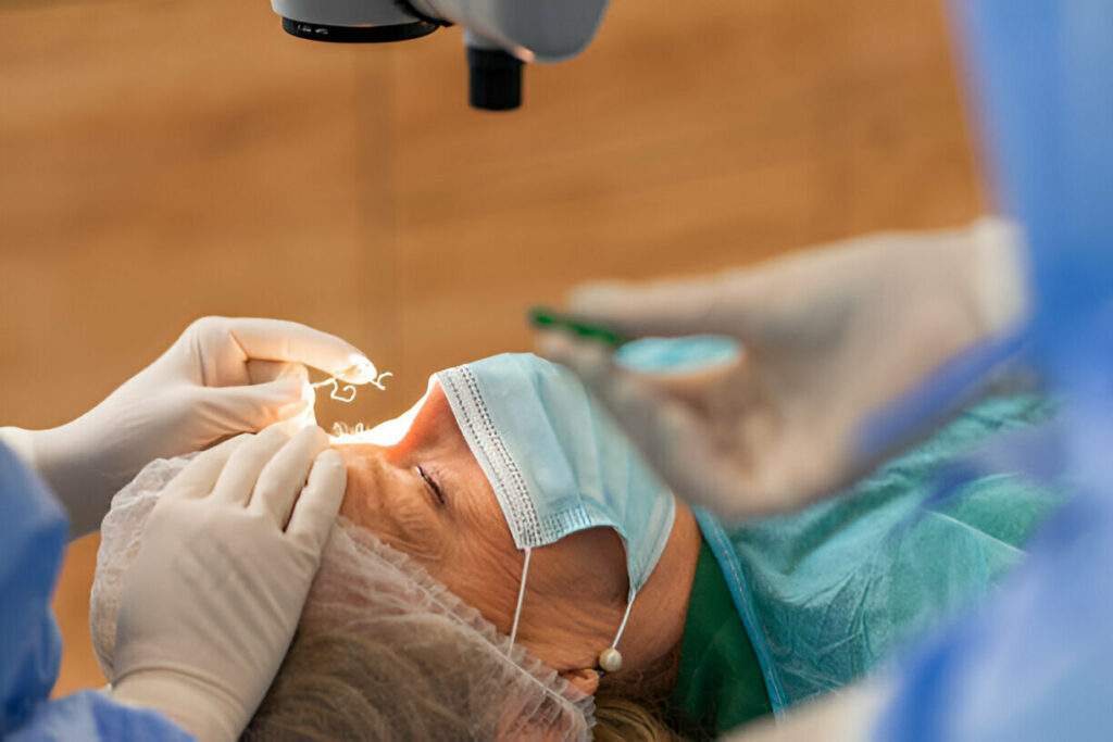
Your doctor will make a small incision and insert an instrument that uses ultrasound energy to fragment and suction away cataract fragments before implanting an artificial lens into your eye.
Infections
Cataract surgery is an increasingly common procedure that replaces the eye’s cloudy natural lens with an artificial one for clearer vision and aiding conditions such as glare and halos. Unfortunately, complications do sometimes arise after cataract removal surgery; however, infections tend to be rarer than other surgeries.
At cataract surgery, a small incision is made on one of the cornea’s sides to enable ultrasound waves to soften and break up cataracts in preparation for removal by suction. Once this process has taken place, an injector tool is then used to implant a new lens, which unfolds before being secured by your surgeon for proper alignment within your eye.
Postoperatively, patients should avoid rubbing or applying pressure to the eye. They may experience mild discomfort, light sensitivity or blurry vision which should dissipate within a few days; drops that reduce inflammation and control eye pressure are typically prescribed as post-op care.
Endophthalmitis, an eye infection caused by bacteria or fungus, can be an extremely serious yet rare side effect of cataract surgery, leading to inflammation inside of the eye, potentially leading to vision loss or blindness.
Endophthalmitis following cataract surgery is rare but potentially lethal, accounting for 0.1% of operations studied. One research paper found that most endophthalmitis infections were caused by Pseudomonas aeruginosa bacteria which were resistant to antibiotics such as gentamicin, amikacin, piperacillin, tetracycline and co-trimoxazole treatments.
Researchers are studying the causes and ways to prevent infection following joint replacement surgery. A shift in surgical techniques or patient selection could potentially lower infection risks.
Posterior Capsular Opacification (PCO)
Cataract surgery often results in improved vision for most patients, though complications from it can include issues like glare and blurriness. A common complication associated with cataract removal surgery is posterior capsular opacification (PCO). After cataract extraction, part of the thin, clear membrane that holds in place the artificial lens may become cloudy or opaque leading to poor quality vision as well as potentially further complications like retinal detachment.
PCO rates have declined greatly thanks to advances in intraocular lenses that actually reduce its incidence. Still, it occurs in about one third of people who undergo cataract surgery due to cells left behind that grow and multiply following cataract removal opacifying their lenses and leading to PCO.
Researchers aimed to identify risk factors for PCO by reviewing data from a retrospective study on 500,872 cataract operations conducted on 373,579 eyes at 41 centers by 2,196 surgeons between 1998 and 2008. A number of factors were discovered that could influence PCO development: age of the patient, their gender and the type of intraocular lens inserted.
Researchers used a dark field microscope to measure whether light scattering in PCO changed after it was fixed with paraformaldehyde (PFA). Light intensity measurements were made for each region of PCO before and after PFA fixation; light intensity averages for all regions were then compared. They discovered no statistically significant difference in light intensity after PFA fixation was applied;
Once your procedure is over, the next step should be identifying what caused opacification in the lens capsule afterward – typically an infection or inflammation – so as to address it effectively and restore clear vision. Failing that, the opacification may persist resulting in further issues with vision; alternatively, your doctor may need to perform laser surgery known as Neodymium:Yttrium-Aluminum-Garnet (Nd:YAG) capsulotomy to eliminate it and restore clear sight.
Dislocation of the Intraocular Lens (IOL)
Cataract surgery is one of the most frequently performed eye procedures by ophthalmologists, and can significantly improve vision for those affected by cataracts. Cataracts occur due to clouding or opacification in your natural crystalline lens of your eye and usually produce blurry or glarey light, difficulty focusing, as well as other symptoms. While certain symptoms of cataracts can be treated using prescription eyeglasses or other remedies, cataract surgery remains the best way of eliminating cataracts entirely.
At cataract surgery, your surgeon makes a small incision on one side of your cornea (the transparent dome-shaped surface covering the front part of your eye) before inserting a probe emitting high-frequency ultrasound waves to break up and soften the center of the old lens. Once softened, this allows them to suction out and replace with an artificial lens known as an intraocular lens (IOL), made out of plastic, silicone or acrylic that helps focus light onto your retina and restore clear sight.
However, if an IOL becomes misalign or dislocated after surgery, it can result in double vision or other visual issues. Although rare, repositioning surgery will usually be available to reposition it back in its proper place.
One study determined that complications during cataract surgery, past uveitis and zonule laxity increase the likelihood of late IOL dislocation; they also noted an increase in time to dislocation as severity of cataract increased.
Your doctor can help lower the risks and benefits associated with cataract surgery by providing information and counseling regarding how best to use medication and avoid activities which strain or strain the eyes. Furthermore, regular follow-up visits should be scheduled so your ophthalmologist can monitor recovery progress and detect early warning signs of complications.
Visual Loss
Cataract surgery has an extremely high success rate and should be safe for most individuals, yet any surgical procedure carries risk and complications can arise from it. Most complications related to cataract surgery are treatable; however if they persist they could lead to vision loss or blindness if severe enough. If you have any concerns it’s essential that they are discussed with your eye doctor immediately.
Posterior Capsular Opacification (PCO), is one of the primary causes of visual loss following cataract surgery. PCO occurs when your lens capsule becomes opaque months or years post-op due to an increase in fluid, usually from an implanted lens that remains behind its capsule wall. A laser procedure called YAG capsulotomy may provide relief.
Eye complications that may follow cataract surgery include droopy eyelids, infections or glaucoma – although these complications are relatively uncommon they should still be reported promptly to your eye care professional if experienced.
Vitreous loss is another serious complication from cataract surgery that may arise. This complication may cause flashes of light, vision floaters and eventual loss of vision. Vitreous loss tends to occur more in individuals who have had both eyes affected with cataracts as well as those who already had preexisting health conditions like extreme nearsightedness before surgery began; additionally it’s more likely to happen after suffering a trauma or unplanned surgeries.
Skilled cataract surgeons who perform high volumes of procedures tend to have lower rates of intraoperative complications like vitreous loss. Furthermore, there are risk stratification systems which help your eye surgeon identify higher-risk patients and counsel them accordingly.