
Medically reviewed by Shaun Larsen, OD – Doctor of Optometry, Southern College of Optometry. Low vision rehabilitation and contact lens specialist.
Last clinically reviewed: July 2026 | Next scheduled review: July 2027 | Reading time: 22 minutes
Editorial standard: Every clinical claim on this page is sourced to a peer-reviewed journal, a national eye institute, or a professional-society clinical guideline. Sources are listed in full at the end of the article. This page is educational and is not a substitute for an examination by an eye care professional.
What this atlas covers
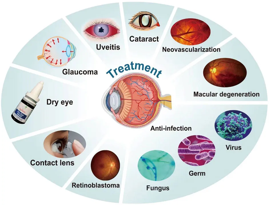
This is a visual reference to the ocular conditions most likely to cause permanent vision loss. Each entry follows the same structure: what physically goes wrong in the eye, what the clinician sees on examination and imaging, what the patient notices, how the condition is staged, and what the current evidence supports as treatment.
Conditions are ordered from the front of the eye backwards – cornea, then lens, then optic nerve, then retina – because that is the order in which an eye examination proceeds and the order in which the accompanying image set is arranged.
A note on the images. Retinal photographs in this guide are illustrative of typical presentations. Disease appearance varies enormously with fundus pigmentation, media clarity, camera field of view, and stage. A photograph cannot diagnose you, and an eye that looks normal in a photograph can still harbour early glaucoma or diabetic change detectable only on OCT. Use these images to understand what your clinician is describing, not to self-assess.
1. Cataract: opacification of the crystalline lens
A cataract is a cloudy condition of the natural lens inside of an eye that affects vision, normally clear lens that becomes clouded over time and causes light rays to scatter, blurring vision. The crystalline lens sits behind the iris and contributes roughly 20 dioptres of the eye’s total focusing power. It is built from tightly ordered crystallin proteins in a cellular architecture that has no blood supply and sheds no cells, so damaged protein accumulates across a lifetime. As those proteins aggregate, the lens scatters and absorbs light rather than transmitting it cleanly.
The three morphological types matter clinically, because they produce different symptoms:
- Nuclear sclerotic cataract – hardening and yellowing of the lens centre. Progresses slowly over years. Characteristically causes a myopic shift, so some patients experience a temporary improvement in reading vision without glasses. This is “second sight”, and it is a sign of progression, not recovery.
- Cortical cataract – wedge-shaped spoke opacities beginning at the lens periphery and advancing toward the visual axis. Strongly associated with diabetes and UV exposure. Produces disproportionate glare because the spokes scatter light obliquely.
- Posterior subcapsular cataract (PSC) – a plaque immediately in front of the posterior lens capsule, directly on the visual axis. Causes symptoms far out of proportion to its size, particularly in bright light and when reading, because pupillary constriction narrows the aperture onto the opacity. PSC is the type most associated with corticosteroid use – inhaled, oral or topical – and with radiation exposure.
Cataracts are graded in research settings using the Lens Opacities Classification System III (LOCS III), which scores nuclear opalescence, nuclear colour, cortical and posterior subcapsular changes against standard photographs. In routine practice, the operative decision is driven by symptoms and functional impairment rather than by grade alone.
Established risk factors: advancing age, cigarette smoking, diabetes mellitus, cumulative UV-B exposure, corticosteroid therapy, prior intraocular surgery or trauma, high myopia, and ionising radiation. Age is not modifiable; most of the rest are.
Symptoms of cataracts include blurry vision, difficulty driving at night due to glare and color changes. People living with cataracts describe their vision as like looking through fogged windows. Although cataracts typically are painless conditions, they can interfere with daily activities.
A critical measurement note: standard Snellen acuity is measured with high-contrast black letters on a bright white chart, which is close to the best-case scenario for a cataractous eye. Patients frequently read 20/30 in the examination lane yet cannot drive at night. Glare testing (brightness acuity testing) and contrast sensitivity measurement expose the deficit that the acuity chart hides. If your vision is worse in the real world than your chart result suggests, ask for these tests specifically.
Treatment. Early cataract is managed optically: an updated spectacle prescription, task lighting directed over the shoulder onto the page, and anti-glare filters. There is no eye drop, supplement or exercise proven to reverse lens opacity.
When symptoms outweigh those measures, phacoemulsification is the standard procedure. A 2–2.8 mm incision is made at the corneal limbus, a circular opening is created in the anterior lens capsule (capsulorhexis), ultrasonic energy emulsifies the lens nucleus, the fragments are aspirated, and a foldable intraocular lens (IOL) is placed inside the retained capsular bag. It is performed under topical or local anaesthesia, typically in under 20 minutes, and is one of the most frequently performed and highest-success operations in medicine.
IOL choice is a genuine decision point. Monofocal lenses give one focal distance with excellent contrast; multifocal and extended-depth-of-focus lenses reduce spectacle dependence at the cost of some contrast sensitivity and a higher rate of halo and glare; toric lenses correct corneal astigmatism. Patients with existing macular disease, glaucoma or corneal irregularity are generally poor candidates for multifocal optics, because those designs spend contrast that a compromised retina cannot afford to lose.
Cataract surgery in eyes with other disease. Removing a cataract restores media clarity; it cannot restore a retina or optic nerve already damaged by AMD, diabetic retinopathy or glaucoma. Where both coexist, surgery is still often worthwhile – it improves the clinician’s view of the retina for monitoring and treatment, and it removes one of two limiting factors – but expectations must be set explicitly beforehand. Where best-corrected acuity remains reduced after surgery, low vision rehabilitation is the appropriate next step, not a second operation.
2. Glaucoma: progressive optic neuropathy
Glaucoma is a group of progressive optic neuropathies characterised by accelerated loss of retinal ganglion cells and their axons, producing a distinctive pattern of optic nerve head excavation and corresponding visual field loss. Raised intraocular pressure (IOP) is the principal modifiable risk factor and the only one treatment currently targets – but it is a risk factor, not the definition.
This distinction is clinically consequential. A substantial proportion of open-angle glaucoma in some populations occurs at IOP that never exceeds the statistically “normal” ceiling of 21 mmHg. This is normal-tension glaucoma, and patients told their pressure is fine are sometimes falsely reassured by exactly the framing the previous version of this page used. Conversely, many people with pressures above 21 mmHg never develop glaucomatous damage at all; that is ocular hypertension, a risk state rather than a disease.
Primary open-angle glaucoma (POAG) is the most common form in Western populations. The drainage angle is anatomically open, but outflow resistance rises within the trabecular meshwork and juxtacanalicular tissue. Onset is insidious, painless, and initially confined to the mid-peripheral field – an arcuate or nasal-step defect that the fellow eye and the brain fill in seamlessly. Patients typically notice nothing until a large fraction of the ganglion cell population is already lost, which is why glaucoma is detected by examination rather than by symptoms.
Primary open-angle glaucoma is one of the most prevalent types, typically manifesting slowly due to blockages in drainage canals of eyes gradually developing over time.
Primary angle-closure glaucoma (PACG) is mechanically different: the peripheral iris obstructs access to the trabecular meshwork. It may be chronic and asymptomatic, or it may present as acute angle closure – a true ocular emergency with severe pain, a red eye, a mid-dilated non-reactive pupil, corneal oedema, haloes around lights, and often nausea and vomiting. Acute angle closure requires same-day treatment. Anyone with these symptoms should attend an emergency department or ophthalmologist immediately; permanent loss can occur within hours.
Secondary glaucomas arise from an identifiable cause: pseudoexfoliation, pigment dispersion, neovascularisation (usually from proliferative diabetic retinopathy or ischaemic vein occlusion), uveitis, trauma, or steroid response.
What clinicians examine and measure:
- Intraocular pressure by Goldmann applanation tonometry, interpreted alongside central corneal thickness, which systematically biases the reading – thin corneas under-read true pressure.
- Optic nerve head appearance: vertical cup-to-disc ratio interpreted relative to disc size, neuroretinal rim contour, rim notching, disc haemorrhage, and peripapillary atrophy. A large physiologic cup in a large disc is normal; a 0.5 cup in a small disc can be pathological.
- Gonioscopy to determine whether the drainage angle is open or occludable – this drives the entire treatment pathway and cannot be inferred from pressure alone.
- Optical coherence tomography quantifying peripapillary retinal nerve fibre layer thickness and macular ganglion cell–inner plexiform layer thickness, both of which detect structural loss before it becomes measurable on visual field testing.
- Standard automated perimetry to map functional loss and, over serial tests, to establish rate of progression – the number that actually determines how aggressive treatment needs to be.
Treatment. Every current therapy works by lowering IOP; none restores lost ganglion cells. The goal is to set a target pressure low enough that the rate of progression will not produce disabling vision loss within the patient’s lifetime, then to verify by serial testing that the rate is in fact controlled.
- Topical medication – prostaglandin analogues (increase uveoscleral outflow, once nightly, first-line for most patients), beta-blockers, alpha-2 agonists, topical and oral carbonic anhydrase inhibitors, and Rho-kinase inhibitors.
- Selective laser trabeculoplasty (SLT) – applies short, low-energy pulses to pigmented trabecular meshwork cells. The LiGHT randomised trial reported that SLT as initial therapy achieved comparable disease control to drops with a lower rate of subsequent glaucoma surgery, and it has moved SLT from a second-line option to a reasonable first-line one. It is repeatable and it removes the adherence problem that undermines drop therapy.
- Laser peripheral iridotomy – for occludable or closed angles; creates an alternative aqueous route through the iris and relieves pupillary block. Prophylactic in narrow angles, urgent in acute closure.
- Minimally invasive glaucoma surgery (MIGS) – trabecular micro-bypass stents, canaloplasty, goniotomy and subconjunctival micro-shunts. Lower risk than traditional filtration surgery, more modest pressure reduction; frequently combined with cataract surgery.
- Filtration surgery – trabeculectomy or glaucoma drainage device implantation, reserved for eyes progressing despite maximal tolerated medical and laser therapy, or presenting with advanced damage.
Adherence is the weak link. Real-world persistence with topical glaucoma therapy is poor, and the disease is silent, so patients receive no feedback from stopping. If cost, side effects, or the physical difficulty of instilling drops is a barrier, say so – laser and surgical alternatives exist specifically for this situation.
3. Age-related macular degeneration (AMD)
Macular degeneration is a disease in which the central part of your retina (known as the macula) deteriorates or breaks down, diminishing or losing function over time. The macula provides fine detail vision necessary for threading needles or reading. It occupies roughly the central 5.5 mm of the retina and contains the highest density of cone photoreceptors in the eye. Its central 0.35 mm, the foveola, contains no rod photoreceptors and no overlying inner retinal layers at all – an anatomical arrangement that maximises resolution and is the reason macular disease is so disproportionately disabling relative to the tissue area involved.
Where the disease actually starts. AMD is a disease of the outer retinal complex: the photoreceptor outer segments, the retinal pigment epithelium (RPE), Bruch’s membrane, and the choriocapillaris. The RPE phagocytoses shed photoreceptor outer-segment discs continuously across a lifetime. When that clearance capacity is exceeded, incompletely digested material accumulates between the RPE and Bruch’s membrane as drusen. Chronic complement-mediated inflammation, oxidative stress and lipid deposition within Bruch’s membrane compound the problem.
Drusen classification predicts risk and is the basis of staging:
- Drupelets (< 63 µm) – normal ageing, not AMD.
- Medium drusen (63–125 µm) – early AMD.
- Large drusen (> 125 µm), or medium drusen with pigmentary abnormality – intermediate AMD. This is the stage at which AREDS2 supplementation has proven benefit.
- Reticular pseudodrusen (subretinal drusenoid deposits) – located above rather than below the RPE, best seen on near-infrared reflectance and OCT, and an independent marker of higher progression risk and impaired dark adaptation.
Dry (non-neovascular) AMD and geographic atrophy
Roughly 85–90% of AMD is non-neovascular. Its advanced form is geographic atrophy (GA): sharply demarcated areas of RPE and photoreceptor loss with visible underlying choroidal vessels. GA typically begins parafoveally and spares the fovea until late, which is why patients often retain reasonable Snellen acuity while being functionally unable to read – the letters they can resolve fall into an atrophic scotoma when they try to track a line of text. Reading speed and microperimetry capture this; acuity does not.
Treatment for dry AMD:
- AREDS2 supplementation for intermediate AMD or advanced AMD in one eye. The formulation is 500 mg vitamin C, 400 IU vitamin E, 10 mg lutein, 2 mg zeaxanthin, 80 mg zinc (as zinc oxide) and 2 mg copper (as cupric oxide). It reduces the risk of progression to advanced AMD by approximately 25%. Two caveats that are widely misunderstood: it does not prevent AMD in people who do not have it, and it does not restore vision already lost. The original AREDS formula contained beta-carotene, which raised lung-cancer risk in smokers and former smokers; lutein and zeaxanthin replaced it, and any product still containing beta-carotene is built on the superseded formula.
- Complement inhibitors for geographic atrophy. Pegcetacoplan (a C3 inhibitor) and avacincaptad pegol (a C5 inhibitor) were approved by the FDA in 2023 and are the first therapies shown to slow GA lesion growth. Their limitations must be stated honestly: they slow anatomical enlargement of the atrophic lesion; the pivotal trials did not demonstrate improvement in visual function, they require continuing intravitreal injections, and reported risks include intraocular inflammation, a raised rate of conversion to neovascular AMD, and rare cases of ischaemic optic neuropathy and retinal vasculitis. Whether the anatomical benefit justifies the burden is a genuine, ongoing debate among retina specialists, and is a decision to make with your own retina specialist rather than from a web page.
- Smoking cessation. Smoking is the strongest modifiable risk factor for AMD, associated with roughly double to quadruple the risk and earlier onset. Cessation reduces risk over subsequent years.
Wet (neovascular) AMD
In neovascular AMD, new vessels grow from the choroid (or, in type 3 lesions, from the retinal circulation) into or beneath the retina. These vessels are structurally incompetent: they leak fluid and blood, distort the photoreceptor mosaic, and ultimately organise into fibrotic scar. Macular neovascularisation is classified by location on OCT and OCT angiography – type 1 (beneath the RPE), type 2 (above the RPE, subretinal), and type 3 (intraretinal angiomatous proliferation).
Wet AMD is the urgent one. New distortion of straight lines (metamorphopsia), a new central grey or dark patch, or sudden central blur warrants examination within days, not at the next routine appointment. Untreated, neovascular AMD can destroy central vision within weeks to months, and vision lost to subretinal fibrosis does not come back. An Amsler grid checked one eye at a time is a reasonable home monitoring tool for at-risk patients – though it is insensitive and a normal grid never rules out progression.
Treatment: intravitreal anti-VEGF therapy, which transformed prognosis in this disease from near-inevitable central vision loss to stabilisation or improvement in the majority of treated eyes. Agents in use include bevacizumab (used off-label, at a small fraction of the cost of the licensed agents), ranibizumab, aflibercept 2 mg, brolucizumab, faricimab (a bispecific antibody targeting both VEGF-A and angiopoietin-2), and high-dose aflibercept 8 mg. The newer agents were developed principally to extend the interval between injections rather than to improve peak acuity outcomes, because treatment burden – not efficacy – is the main driver of real-world undertreatment and the gap between trial and clinic results.
Regimens are individualised: fixed-interval dosing, pro re nata (treat on evidence of activity), or treat-and-extend, in which the interval is lengthened while the macula stays dry and shortened when fluid returns. Photodynamic therapy and thermal laser photocoagulation are now used only in narrow circumstances such as polypoidal choroidal vasculopathy; they are no longer front-line therapy.
Risk factors: age is dominant – prevalence rises steeply after 60. Others include smoking, family history, European ancestry, and common genetic variants in the complement factor H (CFH) and ARMS2/HTRA1 loci. Cardiovascular risk factors and dietary pattern show consistent but more modest associations.
4. Keratoconus: progressive corneal ectasia
Keratoconus is an eye disease that results in the cornea assuming a cone-like shape, leading to blurry and distorted vision and uncorrected astigmatism. Mechanically, it is a progressive thinning and biomechanical weakening of the corneal stroma. Loss of collagen cross-link density and disruption of lamellar architecture allow normal intraocular pressure to bow the weakened tissue forward into a cone, most often inferotemporal to the corneal apex. While usually both eyes are affected, one usually develops it more than the other – marked asymmetry is typical, and a genuinely unilateral case should prompt a search for asymmetric eye rubbing.
The optical consequence is irregular astigmatism: the corneal surface no longer approximates a smooth toric shape, so the aberration cannot be neutralised by a spectacle lens. This is why patients report that new glasses never quite work, that vision is smeared with ghosting and starbursting around lights, and that their prescription changes repeatedly and in inconsistent directions.
Diagnosis is by corneal imaging, not by refraction. Placido-disc topography maps the anterior surface; Scheimpflug tomography (for example Pentacam) additionally maps the posterior surface and a full pachymetric profile, and posterior elevation change is often the earliest detectable abnormality. Key indices are maximum keratometry (Kmax), thinnest pachymetry, the index of surface variance, and posterior elevation at the thinnest point. Serial imaging – not a single scan – establishes progression, conventionally defined by a documented increase in Kmax alongside corroborating change in pachymetry or refraction.
Experts remain uncertain as to the exact cause of keratoconus; however, it tends to run in families and is linked with conditions like hay fever, asthma, excessive eye rubbing and connective tissue disorders like Ehlers-Danlos syndrome. Eye rubbing deserves particular emphasis because it is the one major risk factor a patient controls directly: it is mechanically implicated, dose-related, and strongly associated with atopic disease and with obstructive sleep apnoea. Treating the underlying allergy so that the eyes stop itching is a legitimate part of keratoconus management. Down syndrome carries a markedly elevated prevalence. Onset is usually in the teens or twenties, and progression typically decelerates by the fourth decade as age-related corneal stiffening occurs naturally. Regular eye exams can detect keratoconus early and lead to treatments which could stop its progress; be sure to seek medical advice immediately if any vision changes arise or you experience symptoms like glares, halos or streaking lights.
Treatment splits into two entirely separate objectives, and conflating them is the most common patient misunderstanding:
(1) Halting progression – corneal cross-linking (CXL). Riboflavin is applied to the stroma and activated with ultraviolet-A light, generating reactive oxygen species that form additional covalent bonds between and within collagen fibrils, stiffening the tissue. In the standard epithelium-off Dresden protocol, the central epithelium is debrided (riboflavin cannot cross intact epithelium), riboflavin is instilled, and the cornea is irradiated at 3 mW/cm² for 30 minutes, delivering 5.4 J/cm². Accelerated protocols deliver equivalent total energy at higher irradiance over shorter times. Stromal thickness of at least 400 µm is required to keep UV exposure at the endothelium below its damage threshold. CXL is the only intervention shown to halt progression, and epithelium-off remains the protocol with the strongest long-term evidence. It stabilises the cornea; it is not primarily a vision-improving procedure, though modest flattening and acuity gain are commonly observed. Because it prevents future loss rather than recovering past loss, timing matters: it should be done when progression is documented, not deferred until vision is poor.
(2) Restoring vision – optical correction. In sequence of increasing corneal irregularity: spectacles and soft toric lenses in mild disease; custom soft keratoconus lenses; corneal rigid gas-permeable lenses, which vault the irregular surface with a tear reservoir that neutralises the aberration; piggyback and hybrid designs for comfort; and scleral lenses, which land entirely on the conjunctiva over the sclera, clear the cornea completely, and are now the mainstay for moderate-to-advanced disease. Intrastromal corneal ring segments can regularise the cone in selected eyes. Deep anterior lamellar keratoplasty or penetrating keratoplasty is reserved for corneas with central scarring or intolerance of all contact lens options – a minority of patients, and a proportion that has fallen substantially since CXL became available.
5. Retinal Detachment: a surgical emergency
Retinal detachment occurs when part or all of the neurosensory retina separates from the underlying retinal pigment epithelium, resulting in vision becoming blurry or shadowy and medical attention is needed immediately as this could result in permanent blindness if left untreated. The neurosensory retina and the RPE are apposed but not anatomically fused – they derive from the two walls of the embryonic optic cup and are held together by RPE apical microvilli, interphotoreceptor matrix adhesion, and active subretinal fluid pumping by the RPE. Separate them and the photoreceptors lose the RPE support on which they depend for outer-segment renewal, retinoid recycling and oxygen supply from the choroid. Photoreceptor apoptosis begins within days, which is the reason urgency is measured in hours to days rather than weeks.
The three mechanisms:
- Rhegmatogenous (from the Greek rhegma, a break) – the most common. A full-thickness retinal break allows liquefied vitreous to pass into the subretinal space. Most breaks arise during posterior vitreous detachment, when the vitreous separates from the retina and exerts traction at points of firm adhesion. Risk factors: age, high myopia, prior cataract surgery, blunt trauma, lattice degeneration, and a personal or family history of detachment.
- Tractional – fibrovascular membrane contraction mechanically pulls the retina forward without any break. The dominant cause is proliferative diabetic retinopathy; also seen in sickle-cell retinopathy, retinopathy of prematurity, and penetrating trauma.
- Exudative (serous) – fluid accumulates subretinally from a leaking source with no break and no traction. Causes include choroidal tumours, posterior scleritis, Vogt–Koyanagi–Harada disease, central serous chorioretinopathy, and severe hypertensive or pre-eclamptic choroidopathy. The detachment characteristically shifts with head position. Treatment is directed at the underlying disease, not at the retina – which is precisely why the mechanism must be correctly identified before surgery is contemplated.
Retinal tears allow watery fluid from vitreous space to pass through and collect under retina, isolating it from oxygen and nutrients provided to its layers under it, leading to retinal detachments; three main types are: rhegmatogenous, tractional and exudative.
Signs of retinal detachment include an opaque curtain-like shadow that begins in the center of your vision and may progress outward, eventually covering all fields of view. In practice the shadow more often begins in the periphery and advances centrally, and because the retinal image is inverted, a superior field shadow corresponds to an inferior retinal detachment. Sometimes accompanied by sudden appearance of floaters – although normal for ageing processes; their sudden presence can be an indicator of detached retinas.
The symptom triad that should trigger same-day examination: a sudden shower of new floaters (especially many small dark specks, which may represent pigment or blood); photopsia – brief arcing flashes in the temporal periphery, most obvious in dim conditions; and a progressive peripheral shadow or curtain. Central vision loss means the macula has detached, and macular status is the single strongest predictor of final acuity. A macula-on detachment operated promptly frequently retains excellent vision; once the macula detaches, outcomes fall substantially and continue to decline with each day of delay. If you have these symptoms, treat it as an emergency.
Repair. Retinal breaks without detachment can often be sealed in clinic by laser retinopexy or cryotherapy. Established detachment requires surgery: pneumatic retinopexy (intravitreal gas bubble plus retinopexy, with strict postoperative positioning) for selected superior breaks; scleral buckling, in which a silicone band indents the sclera to relieve vitreous traction – still favoured in younger phakic patients; and pars plana vitrectomy, which removes the vitreous, drains subretinal fluid, treats the breaks and tamponades with gas or silicone oil. Primary reattachment succeeds in the large majority of cases; the principal cause of failure is proliferative vitreoretinopathy, a scarring response that recontracts the retina.
6. Diabetic Retinopathy: microvascular and neurodegenerative retinal disease
Diabetic Retinopathy occurs when chronic hyperglycaemia damages the small blood vessels that nourish the retina (an inner layer at the back of the eye). It is the leading cause of blindness in working-age adults (roughly 20–74 years) in the United States and other high-income countries – an important precision, since AMD is the leading cause overall once older adults are included.
Pathophysiology. Sustained hyperglycaemia drives polyol-pathway flux, advanced glycation end-product formation, protein kinase C activation and oxidative stress. Retinal capillary pericytes are lost, basement membrane thickens, and endothelial tight junctions fail – breaking the inner blood–retinal barrier. Capillaries occlude, producing patches of retinal non-perfusion; the ischaemic retina upregulates vascular endothelial growth factor; and VEGF drives both the vascular leakage that causes macular oedema and the neovascularisation that defines proliferative disease. Retinal neurodegeneration is now recognised to begin before visible vascular lesions appear, which is why some patients report subtle contrast and colour deficits while their fundus still looks unremarkable.
At first, in what’s known as non-proliferative diabetic retinopathy (NPDR), retinal blood vessels either leak fluid or close entirely, creating microaneurysms or localized sac-like swellings in the retina. Over time, due to decreased oxygen and blood supply, they begin growing fragile new blood vessels that bleed into the clear gel-like vitreous humour filling the center of your eyeball.
Staging (ETDRS-derived international clinical classification):
- No apparent retinopathy – no abnormalities.
- Mild NPDR – microaneurysms only.
- Moderate NPDR – more than microaneurysms but less than severe.
- Severe NPDR – defined by the 4-2-1 rule: extensive intraretinal haemorrhages in all 4 quadrants, or definite venous beading in 2 or more quadrants, or prominent intraretinal microvascular abnormalities (IRMA) in 1 or more quadrant, with no neovascularisation. This stage carries a high one-year rate of progression to proliferative disease and changes the monitoring interval sharply.
- Proliferative DR (PDR) – neovascularisation of the disc, retina or iris, and/or vitreous or preretinal haemorrhage. Complications: tractional retinal detachment, vitreous haemorrhage, and neovascular glaucoma when new vessels obstruct the drainage angle.
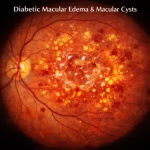
Diabetic macular oedema (DMO/DME) is a separate axis from the staging scale above and can occur at any stage. Fluid accumulates in the macula from blood–retinal barrier breakdown, forming intraretinal cystoid spaces and subretinal fluid. It is the most common cause of vision loss in diabetes. Centre-involving DMO – oedema within the central 1 mm subfield on OCT – is what determines whether treatment is indicated. Note that macular oedema does not cause floaters; floaters in diabetes usually indicate vitreous haemorrhage, which is a different complication requiring different management.
Detection. Dilated fundus examination is the traditional standard, but OCT is what actually detects and quantifies macular oedema – it resolves fluid invisible on clinical examination and provides the reproducible central subfield thickness measurement used to guide retreatment. Widefield imaging and OCT angiography map peripheral non-perfusion without dye. FDA-cleared autonomous AI screening systems now grade diabetic retinopathy from fundus photographs in primary care, and are appropriate for screening but not for managing established disease.
Treatment.
- Systemic control is the foundation and the only intervention that modifies the underlying disease. The DCCT and UKPDS trials established that intensive glycaemic control substantially reduces the incidence and progression of retinopathy; blood pressure and lipid control add further benefit. One caution worth knowing: rapid correction of long-standing severe hyperglycaemia can cause transient early worsening of retinopathy, so patients starting intensive therapy with existing significant disease need closer ophthalmic monitoring – this is a reason for surveillance, never a reason to avoid controlling glucose.
- Intravitreal anti-VEGF therapy is first-line for centre-involving DMO with reduced acuity, and also regresses neovascularisation in proliferative disease. Corticosteroid implants (dexamethasone, fluocinolone acetonide) are alternatives, particularly in pseudophakic eyes or where anti-VEGF response is inadequate, at the cost of cataract progression and IOP elevation.
- Focal/grid laser photocoagulation retains a role for non-centre-involving oedema with discrete leaking microaneurysms, but has been displaced by anti-VEGF as first-line for centre-involving disease.
- Panretinal photocoagulation (PRP) remains the definitive treatment for proliferative disease. By ablating ischaemic peripheral retina it lowers VEGF production and causes neovascular regression. It costs peripheral field and night vision – a real trade the patient should be told about – but it durably prevents severe vision loss, and unlike anti-VEGF it does not depend on the patient returning for indefinite repeat injections.
- Pars plana vitrectomy for non-clearing vitreous haemorrhage or tractional detachment threatening the macula.
Screening intervals (American Academy of Ophthalmology / American Diabetes Association): type 1 diabetes – first dilated examination 5 years after diagnosis; type 2 diabetes – at diagnosis, because retinopathy is frequently already present; pregnancy with pre-existing diabetes – before conception or in the first trimester, then per findings, as pregnancy accelerates progression. Gestational diabetes alone does not require examination during pregnancy. Thereafter at least annually, and considerably more often with established disease. Because early and even moderately advanced diabetic retinopathy is entirely asymptomatic, waiting for symptoms means presenting at the stage where damage is already irreversible.
7. Retinal vein occlusion
Occlusion of the central retinal vein or a branch vein produces sudden, painless, usually unilateral vision loss. Branch retinal vein occlusion typically occurs at an arteriovenous crossing where a thickened arteriole compresses the vein; central retinal vein occlusion occurs at or posterior to the lamina cribrosa. The fundus shows tortuous dilated veins, flame and dot-blot haemorrhages in the drainage territory, cotton-wool spots and macular oedema.
RVO is a systemic vascular event presenting in the eye. Hypertension, hyperlipidaemia, diabetes and smoking are the principal associations, and open-angle glaucoma is an independent ocular risk factor. In patients under 50, thrombophilia and inflammatory screening is warranted. Vision loss occurs through macular oedema and through ischaemia; ischaemic CRVO additionally risks neovascular glaucoma, classically around three months. Treatment is intravitreal anti-VEGF for macular oedema, corticosteroid implants as an alternative, and panretinal photocoagulation for neovascular complications – alongside cardiovascular risk-factor management, which is where the patient’s longer-term prognosis actually lies.
8. Retinitis pigmentosa and inherited retinal disease
Retinitis pigmentosa is a group of inherited rod–cone dystrophies. Rod photoreceptors degenerate first, producing night blindness and progressive concentric peripheral field constriction; central acuity is often preserved until late, leaving patients with good chart acuity but severely restricted mobility – a dissociation that makes acuity-based definitions of impairment particularly misleading here. Classic signs are bone-spicule pigmentation in the mid-periphery, attenuated retinal arterioles, and a waxy pale disc. Full-field electroretinography shows reduced and delayed responses, often long before fundus changes are obvious.
Inheritance may be autosomal dominant, autosomal recessive, X-linked or mitochondrial, with mutations identified in more than 100 genes; RP may also occur syndromically, as in Usher syndrome (with hearing loss) and Bardet–Biedl syndrome. Genetic testing has become clinically actionable rather than merely academic: voretigene neparvovec is an approved gene therapy for biallelic RPE65-mediated retinal dystrophy, and genotype determines eligibility for a growing set of trials. Management otherwise centres on low vision rehabilitation, orientation and mobility training, UV protection, and treatment of cystoid macular oedema where present.
9. Uveitis
Intraocular inflammation, classified anatomically as anterior (iritis, iridocyclitis), intermediate, posterior, or panuveitis. Anterior uveitis presents with pain, photophobia, redness concentrated around the limbus, and cells and flare in the anterior chamber; posterior uveitis is often painless and presents with floaters and blurring. Causes include autoimmune disease (notably HLA-B27-associated spondyloarthropathy, sarcoidosis, Behçet disease, juvenile idiopathic arthritis), infection (toxoplasmosis, herpetic disease, tuberculosis, syphilis), and masquerade syndromes including intraocular lymphoma.
Uveitis matters on a list like this because it is a leading cause of preventable blindness in working-age adults, and because its complications – cystoid macular oedema, secondary glaucoma, cataract, and retinal detachment – do the damage rather than the inflammation itself. Uveitis associated with juvenile idiopathic arthritis is characteristically asymptomatic and white-eyed in children, which is why formal screening schedules exist for that group.
10. Dry eye disease
Included because it is by a wide margin the most common cause of the visual complaints that bring people to search for eye disease information, and because it is frequently dismissed. The TFOS DEWS II definition frames it as a multifactorial disease of the ocular surface characterised by loss of tear-film homeostasis, with tear-film instability and hyperosmolarity, ocular surface inflammation and damage, and neurosensory abnormality as aetiological components.
The two overlapping subtypes are aqueous-deficient (reduced lacrimal secretion; associated with Sjögren syndrome and many systemic medications) and evaporative (predominantly meibomian gland dysfunction). Symptoms include burning, grittiness, paradoxical reflex tearing, and – relevant here – fluctuating blurred vision that clears momentarily on blinking, because the tear film is the eye’s first refracting surface. Management is stepwise: preservative-free lubricants, lid hygiene and warm compresses, omega-3 supplementation, topical anti-inflammatories (ciclosporin, lifitegrast, short-course corticosteroid), punctal occlusion, autologous serum drops, and intense pulsed light or thermal pulsation for meibomian disease.
How these images are captured
Understanding the modality helps you interpret what you are shown in clinic:
- Colour fundus photography – the familiar orange retinal view. Documents drusen, haemorrhages, exudates, disc appearance. Widefield and ultra-widefield systems capture peripheral pathology missed by standard 45° fields.
- Optical coherence tomography (OCT) – low-coherence interferometry producing micron-resolution cross-sections of the retinal layers. This is the workhorse of modern retinal practice: it detects fluid, quantifies macular thickness, measures nerve fibre layer, and drives most retreatment decisions.
- OCT angiography (OCTA) – maps blood flow by detecting decorrelation between sequential scans. Visualises the retinal capillary plexuses and macular neovascularisation without dye injection.
- Fluorescein and indocyanine green angiography – dye-based; still the reference standard for characterising leakage, capillary non-perfusion, and choroidal vascular disease.
- Fundus autofluorescence (FAF) – images lipofuscin in the RPE. The definitive method for delineating and measuring geographic atrophy.
- Corneal topography and Scheimpflug tomography – anterior segment surface and thickness mapping; the basis of keratoconus diagnosis.
When to seek care urgently
Same day – emergency: sudden painless loss of vision in one eye; a curtain or shadow moving across the field; a sudden shower of floaters with flashing lights; severe eye pain with a red eye, haloes, nausea and vomiting; sudden double vision; any eye injury or chemical splash; new vision loss with headache, scalp tenderness or jaw pain in anyone over 50 (possible giant cell arteritis – a sight- and life-threatening emergency).
Within days: new distortion of straight lines; a new central grey or dark patch; sudden onset of floaters without flashes; progressive blurring over days.
Routine: gradual blurring over months, glare or haloes at night, difficulty reading small print, changes in colour perception.
Living with permanent vision loss
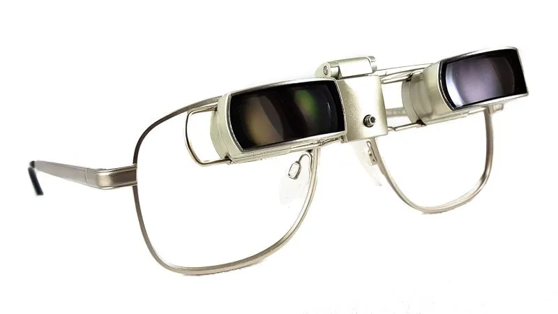
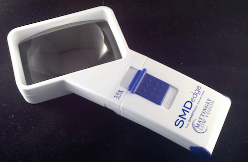
Several conditions on this list can leave irreversible impairment even with optimal treatment. That is the point at which low vision rehabilitation becomes the relevant intervention – and it is chronically under-referred. A low vision assessment measures what remains rather than what is lost: acuity at working distance, contrast sensitivity, visual field, glare tolerance and reading performance. From that, a clinician can prescribe magnification calculated to the patient’s actual reading requirement, task lighting, contrast enhancement, electronic magnification, bioptic telescope systems, eccentric viewing training for central scotoma, and orientation and mobility instruction.
If you have been told nothing further can be done for your eye disease, that statement is about the disease, not about your function. Those are different questions, and the second one usually has answers.
Sources and Medical References
1. Age-Related Eye Disease Study 2 Research Group. Lutein + zeaxanthin and omega-3 fatty acids for age-related macular degeneration: the AREDS2 randomized clinical trial. JAMA. 2013;309(19):2005–2015.
2. National Eye Institute. AREDS/AREDS2 clinical trials – about AREDS and AREDS2. nei.nih.gov.
3. Chew EY, et al. Long-term outcomes of adding lutein/zeaxanthin and omega-3 fatty acids to the AREDS supplements: AREDS2 report 28. JAMA Ophthalmol. 2022;140(7):692–698.
4. Heier JS, Lad EM, Holz FG, et al. Pegcetacoplan for the treatment of geographic atrophy secondary to age-related macular degeneration (OAKS and DERBY): two multicentre, randomised, double-masked, sham-controlled, phase 3 trials. Lancet. 2023;402(10411):1434–1448.
5. American Academy of Ophthalmology. What to know about Syfovre and Izervay for geographic atrophy. aao.org.
6. Gale JD, et al. Five-year GALE extension outcomes for pegcetacoplan in geographic atrophy. Apellis Pharmaceuticals; November 2025.
7. Gale MJ, Wollensak G, Spoerl E, Seiler T. Riboflavin/ultraviolet-A-induced collagen crosslinking for the treatment of keratoconus. Am J Ophthalmol. 2003;135(5):620–627.
8. American Academy of Ophthalmology. Safety and efficacy of epithelium-off corneal collagen cross-linking for the treatment of corneal ectasia: an Ophthalmic Technology Assessment. Ophthalmology. 2024.
9. Gordon MO, Kass MA, et al. The Ocular Hypertension Treatment Study: baseline factors that predict the onset of primary open-angle glaucoma. Arch Ophthalmol. 2002;120(6):714–720.
10. Gazzard G, Konstantakopoulou E, Garway-Heath D, et al. Selective laser trabeculoplasty versus eye drops for first-line treatment of ocular hypertension and glaucoma (LiGHT): a multicentre randomised controlled trial. Lancet. 2019;393(10180):1505–1516.
11. Early Treatment Diabetic Retinopathy Study Research Group. Grading diabetic retinopathy from stereoscopic color fundus photographs – ETDRS report number 10. Ophthalmology. 1991;98(5 Suppl):786–806.
12. Wilkinson CP, Ferris FL, Klein RE, et al. Proposed international clinical diabetic retinopathy and diabetic macular edema disease severity scales. Ophthalmology. 2003;110(9):1677–1682.
13. Diabetes Control and Complications Trial Research Group. The effect of intensive treatment of diabetes on the development and progression of long-term complications in insulin-dependent diabetes mellitus. N Engl J Med. 1993;329(14):977–986.
14. American Academy of Ophthalmology. Diabetic Retinopathy Preferred Practice Pattern. aao.org.
15. Craig JP, Nichols KK, Akpek EK, et al. TFOS DEWS II definition and classification report. Ocul Surf. 2017;15(3):276–283.
16. Verbakel SK, van Huet RAC, Boon CJF, et al. Non-syndromic retinitis pigmentosa. Prog Retin Eye Res. 2018;66:157–186.
17. World Health Organization. International Classification of Diseases, 11th Revision – vision impairment including blindness. who.int.






 Shop Best Low-Vision Aids with FREE Doctor Consultation.